Header Molecular Tumor Diagnostics

MALDI Imaging Unit – Targeting the protein landscape of cancer

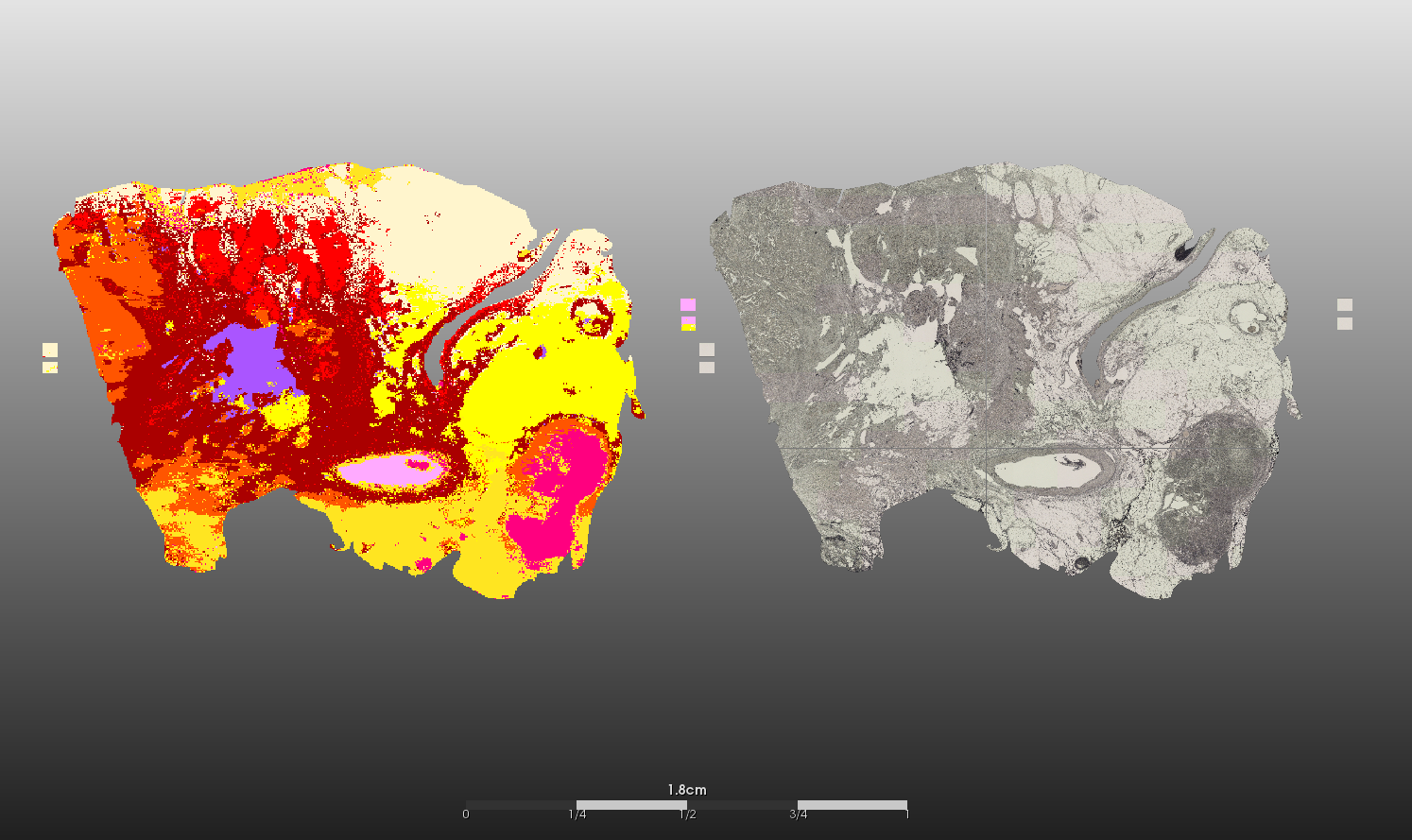
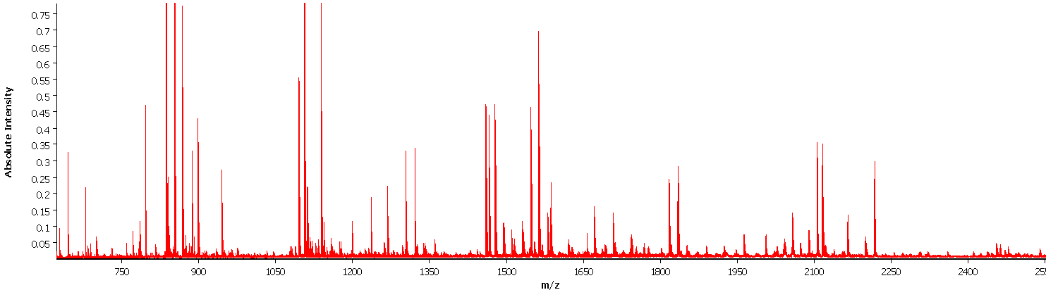
Matrix-associated laser desorption/ionization imaging (MALDI imaging) combines two major fields of bioanalytical methods: molecule analysis using time-of-flight mass spectrometry and the macroscopic anatomy of the tissue compartments. Regions of interest (ROI) of human cancer tissue slides defined by pathologists are translated into mass spectra patterns and can be used for subsequent biostatistical analyses of protein, peptide or lipid composition and quantification. In addition to the knowledge gained from differing mass spectra in e.g. tumor versus normal tissue areas, morphologically indistinguishable tumors can be subdivided and analyzed for molecule-dependent heterogeneity through segmentation of mass intensities at a very high resolution (down to 5x5 micrometers).

In addition to the significance of conventional MALDI analysis for routine diagnostics and for medical research projects using experimental setups, the MALDI Imaging Unit researches bioinformatic workflows and deep learning pipelines to utilize big data e.g. for automation purposes.

Figure 1: MALDI Imaging instrument. Rapflex MALDI Tissuetyper (Bruker Daltonik GmbH) to perform MALDI Imaging analyzation based on time of flight molecule mass detection.

Figure 1: MALDI Imaging instrument. Rapflex MALDI Tissuetyper (Bruker Daltonik GmbH) to perform MALDI Imaging analyzation based on time of flight molecule mass detection.

Figure 2: Visualization of mass spectra pattern on tissue slides via segmentation. A pancreatic tumor sample was analyzed by MALDI imaging. Left side: Visualization of mass spectra as segmentation map based on trained regions of interest of specified tissue compartments. Right side: Grayscale image scan of the same tissue. Red: pancreatic tissue, orange: tumor tissue, dark red: stroma, yellow: fatty tissue, pink: lymph node, lilac/rose: background.

Research focus

  • Tumor classification of rare tumor types
  • Segmentation of (intra-)tumor heterogeneity
  • Lipid, protein and peptide mass spectra identification of novel tumor subtypes
  • Generation of an ever-growing database of tumor vs normal tissue mass spec data
  • MALDI segmentation pipelines based on deep learning
  • 3D MALDI imaging approaches

Sample types for MALDI imaging:

  • Fresh frozen tissue
  • Formalin-fixed paraffin-embedded (FFPE) tissue
  • Samples with and without on-slide enzymatic digestion
  • Embedded organoids, spheres or cell culture blocks

Technical information:

  • Positive and negative flight mode detected by linear or reflector detector
  • Mass range (positive mode e.g. peptides: 600-3200 m/z, proteins 1000-500,000 m/z)
  • Scan range: 5x5µm to 150x150µm resulting field size
  • Automated batch run option
  • External calibration and quality controls provided
  • Big data storage (data files from 20 to 100GB)
Figure 3: Mass spectra of tumor region.  Example of  one mass spectra pattern in a range of 600-3200 m/z of  a tumor region (FFPE sample).

Figure 3: Mass spectra of tumor region. Example of one mass spectra pattern in a range of 600-3200 m/z of a tumor region (FFPE sample).

Coordination

Dr. rer. med. Pia Hönscheid
Group leader
E-Mail: pia.hoenscheid(at)uniklinikum-dresden.de
Phone: +49 (0)351 45813038

Director

Prof. Dr. med. Gustavo Baretton
Director of Institute of Pathology Dresden
E-Mail: Gustavo.Baretton(at)uniklinikum-dresden.de
Phone: +49 (0)351 4583000

Team

Christian Sperling
Medical Technical Assistant
E-Mail: Christian.Sperling(at)uniklinikum-dresden.de
Phone: +49 (0)351 458-13009

Maxime le Floch  
MD Student
E-Mail: maxime.lefloch(at)medforum-dresden.de   
Phone: +49 (0)351 458-3009

Maximilian Weiss
Physician in further education
E-Mail: Maximilian.Weiss(at)ukdd.de 
Phone: +49 (0)351 458-5266